


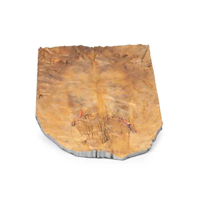


3D Printed Internal Abdominal Wall
This 3D model captures the internal surface of the anterior abdominal wall, a region oftentimes removed or damaged
during dissection (and complimenting our A8 abdominal specimen where the anterior wall has been removed). The
parietal peritoneum has been removed from the internal surface of the specimen in order to more clearly demonstrate
the relationships of the anterior abdominal muscle fibres and connective tissue structures as they converge on the
midline. On the margins of the specimen, particularly superiorly, the horizontally-oriented transversus abdominus
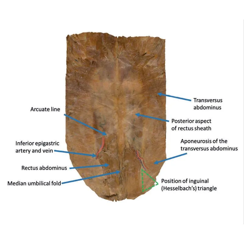
muscle fibres can be seen converging towards their aponeurosis (tendon sheet). In the inferior 1/3 of the model, we
can see the termination of the posterior aspect of the aponeurosis forming the arcuate line. This marks the location
where the aponeurosis changes its orientation relative to the rectus abdominus muscle (visible on either side of the
midline); above the arcuate line the aponeurosis of the transversus abdominus muscle is evenly divided around the
rectus abdominus muscle, while below the arcuate line all aponeurotic fibres pass anterior relative to the rectus
abdominus.
At this point, we can observe the inferior epigastric arteries (and accompanying veins) passing
superiorly from their origins from the external iliac arteries and veins to pass into the anterior abdominal wall
tissues. On the right side of the model we can appreciate how the orientation of the inferior epigastric artery
relative to the fibres of the rectus abdominus muscle define the apex of the inguinal (Hesselbach’s) triangle
(missing only the base formed by the inguinal ligament, not present in this specimen). This region lateral to the
inferior epigastric artery is a frequent site of direct hernias (which can be appreciated on the A8 abdomen model)
given the relative weakness of the wall inferior to the arcuate line and lateral to the margin of the rectus
abdominus muscle. In the midline, and dividing the two halves of the rectus abdominus muscle, is part of the median
abdominal ligament – a draped fold of the parietal peritoneum that covers the urachus, a fibrous embryological
remnant of the allantois, which extends from the bladder into the umbilical cord.
GTSimulators by Global Technologies
Erler Zimmer Authorized Dealer


